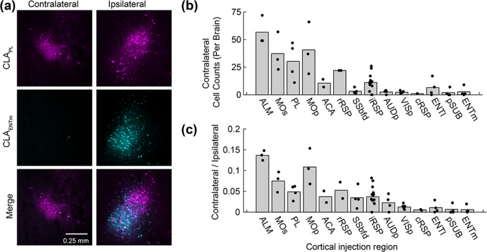
FIGURE 6.

Contralateral projecting claustrum neurons innervate frontal midline cortex but not the temporal lobe. (a) A representative experiment showing labeling in the contralateral and ipsilateral claustrum following retrograde tracer injections into PL (magenta, AAV2‐retro‐GFP) and ENTm (cyan, AAV2‐retro‐tdtomato). Note the absence of claustrum neurons in the contralateral hemisphere, following retrograde tracer deposited into the ENTm. (b) The total number of neurons counted in 5–6 slices in the contralateral hemisphere for each cortical region injection. (c) The ratio of contralateral/ipsilateral labeling in the claustrum for each cortical injection region. Contralateral labeled was mainly found in the case of claustrocortical inputs to motor related regions and was less prominent in the case of injections into sensory cortex and temporal lobe. Each point represents one mouse, and the bar plot shows the mean [Color figure can be viewed at wileyonlinelibrary.com]
