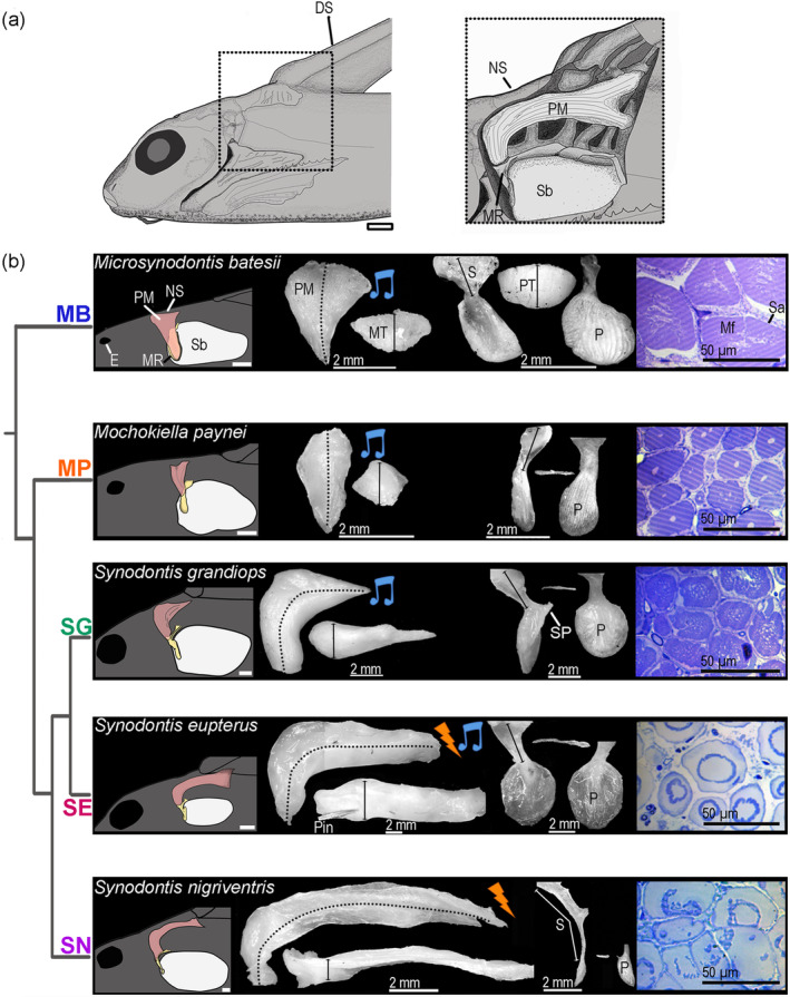
FIGURE 5.

Morphology of the elastic spring apparatus of five mochokid catfish. (a) Schematic of the head and anterior trunk of a Synodontis eupterus in a left lateral view. The position of the elastic spring apparatus (ESA) is outlined in the box. The right part shows a magnified view of the ESA. (b) Drawing of the ESA and the swim bladder (far left); lateral and dorsal views of the protractor muscle (middle left); lateral, ventral, and inner views of the MR (middle right); and transverse section of the protractor muscle (far right) for each of the five mochokid species placed according to their phylogenetic relationship which was inferred from Day et al. (2013). Pictures of the protractor muscle and MR were standardized along the y axis. DS, dorsal spine; E, eye; Mf, myofibrils; MR, Müllerian ramus; MT, muscle thickness; NS, nuchal shield; P, plate; PT, plate thickness; S, stem of the MR; Sa, sarcoplasm; Sb, swim bladder; SP, stem process. Scale bars for far left and two middle panels: 2 mm. Scale bar for far right panels: 50 μm [Color figure can be viewed at wileyonlinelibrary.com]
