
Figure 5.
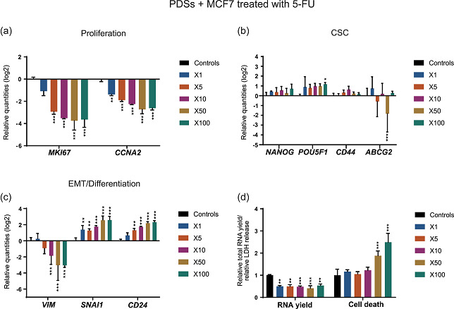
Treatment with 5‐FU enhances the CSC low proliferative phenotype in PDSs cultured with MCF7 cells. Modulation of the expression of genes related to (a) proliferation, (b) CSC and (c) EMT/differentiation at increasing concentrations of 5‐FU indicated as fold‐change respect to the IC50 in 2D cultures. Data is relative to gene expression in untreated PDSs (log2). (d) 5‐FU effect on the total RNA yield and LDH release, as surrogate measurements for cell count and cell death, respectively, quantified at the same drug concentrations. Relative quantities to untreated PDSs are represented. Mean ± SD is shown, n = 3. Significant differences to the untreated controls are stated (*p < .05, **p < .01, and ***p < .001). 5‐FU, 5‐fluorouracil; CSC, cancer stem cell; EMT, epithelial–mesenchymal transition; LDH, lactate dehydrogenase; PDS, patient‐derived scaffold