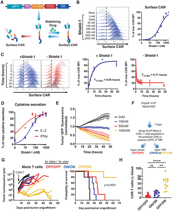
Figure 1: A GD2-targeting CAR modified with a destabilizing domain (DD) exhibits drug-dependent control of expression, function, and tonic CAR signaling.
A) Schematic depicting drug-dependent control of DD CAR fusion protein. B) Flow cytometric analysis of GD2.28ζ.FKBP CAR surface expression at increasing concentrations of shield-1. C) Flow cytometric analysis of ON/OFF kinetics of GD2.28ζ.FKBP CAR surface expression at indicated time points after addition or removal shield-1. D) IL-2 and IFNγ secretion of GD2.28ζ.FKBP CAR-T cells pretreated with indicated concentrations of shield-1 16 hours prior to co-culture with Nalm6-GD2 leukemia. E) Cytotoxicity of GD2.28ζ.FKBP CAR-T cells treated as in (D) against Nalm6-GD2-GFP leukemia (1:2 E:T, normalized to t=0). Error bars represent mean ± SD of triplicate wells. Representative donor from 3 donors. F-H) 2x10⁶ GD2.28ζ.ecDHFR CAR-T cells expanded in the presence or absence of trimethoprim (TMP) for 15 days in vitro (F) were infused IV in NSG mice 7 days post-engraftment of 1x10⁶ Nalm6-GD2 leukemia cells. Mice were dosed 6 days per week with vehicle (water, OFF/OFF) or 200mg/kg TMP (ON/ON and OFF/ON). (G) Quantification of tumor growth by bioluminescent imaging (right) and survival (left) (p<0.0001 log-rank Mantel-Cox test). Representative experiment from 3 independent experiments (n=5 mice/group). (H) Detection of CAR-T cells in peripheral blood sampled on day 28 post-engraftment by flow cytometry after anti-human CD45 staining (n=10 mice/group from 2 independent experiments). B-C show histograms from one representative donor (n=3 donors). Curves in B-D show mean ± SEM from 3 donors. Statistics: G log-rank Mantel-Cox test, H Kruskal-Wallis and Dunn’s multiple comparisons test. **, p<0.01; ****p<0.0001; ns, p>0.05

