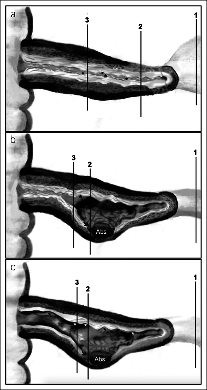
Figure 1.

Schematic representation of anatomical locations of the 3 per-protocol sections for histopathological study, obtained from formalin-fixed and paraffin-embedded blocks of ileal resection surgical specimens (panels a to c). 1—Proximal ileal margin; 2—most affected area; and 3—inflamed area. (a) Schematic surgical specimen with strictures. (b) Schematic surgical specimen with fistulas, fissures, and/or deep ulcers and stricture. (c) Schematic surgical specimen with fistulas, fissures, and/or deep ulcers only. Abs—abscess; asterisks—superficial ulcers; arrow—deep ulcer (beyond submucosa). 1Inflammation (1–3) and fibrosis (0–2) scoring: Higher scores indicate more severe inflammation and fibrosis, respectively (23).
