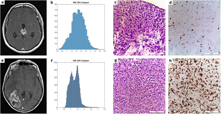
Fig 1. MRI, ADC histogram and histopathological findings in patients with HGG.
Fig 1 shows typical MRI images, the inherent tumor volume ADC histogram as well as H&E staining and Ki-67 immunohistochemistry of a grade III (a-d) and a grade IV glioma (e-h). The first image of the upper case displays a T1 weighted turbo-spin-echo (TSE) sequence (after intravenous application of a gadolinium-based contrast medium) of a grade III astrocytoma, involving the right and left thalamus as well as the aqueduct with consecutive hydrocephalus (a). The first image of the lower case illustrates a contrast enhanced T1 weighted TSE sequence of a grade IV glioblastoma of the right occipital and the adjacent temporal lobe with marked perifocal edema and mass effect (e). Each MRI example is followed by the corresponding ADC histogram (b, f; x-axis: ADC values in incremental order, y-axis: number of voxels), the H&E staining and the Ki-67 immunohistochemistry on the right side (c-d, g-h). A proliferation index of 12% was calculated for the anaplastic astrocytoma and a proliferation index of 80% for the glioblastoma.

