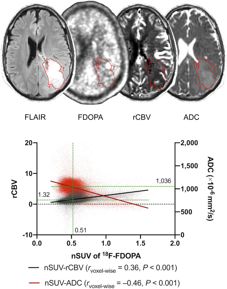
FIGURE 1.
Example of postprocessing and segmentation in 36-y-old man with treatment-naïve WHO grade IV, IDHwt, MGMT-unmethylated, and EGFR-amplified glioblastoma. ROIs of FLAIR hyperintense region are overlaid on 18F-FDOPA, rCBV, and ADC maps. A scatterplot extracted from ROI is shown with rvoxelwise between nSUV and rCBV or ADC. Median nSUV, rCBV, and ADC (green lines) are also shown.

