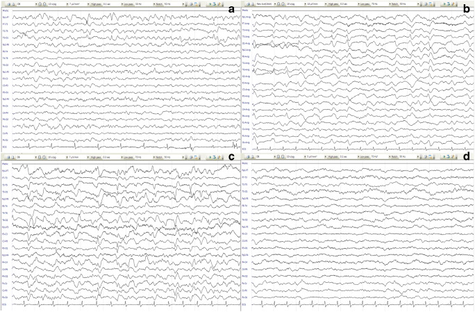
Fig. 2.
a There was very frequent rhythmic (more than 2.5 Hz) left temporal sharp compatible with EEG ictal pattern (starting from: F7> T3, T5)*. b, c There was no posterior dominant rhythm. There was continuous left temporal sharp/periodic lateralized epileptiform discharge (PLED)/EEG ictal pattern (maximum potential: T3, T5) and independent frequent right occipital sharp/PLED (maximum potentialO2)* and a continuous right temporo-occipital rhythmic and irregular delta slowing (maximum potential: O2, T6) which we assumed compatible with focal nonconvulsive status epilepticus. d After tapering the anesthetics, the background rhythm consisted of a low amplitude irregular delta, theta activity without epileptiform discharges. *F frontal, T temporal, O occipital

