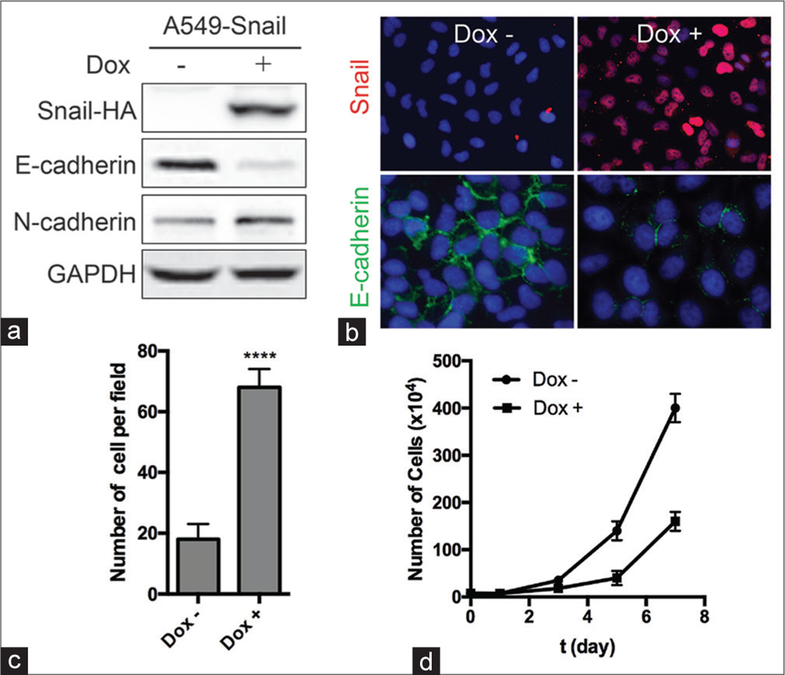
Figure 1:
Overexpression of Snail induces the epithelial–mesenchymal transition in A549 cells. A549‑Snail cells, engineered to stably express Snail‑HA under the control of a doxycycline (Dox)‑inducible promoter, were grown in the absence or presence of 2 μg/ml Dox for 2 days. (a) The levels of Snail‑HA, E‑cadherin, N‑cadherin, and GAPDH proteins were measured using Western blotting. (b) The levels of Snail‑HA (red) and E‑cadherin (green) were visualized using immunohistochemistry, and the nuclei were visualized using 4′,6‑diamidino‑2‑phenylindole staining (blue). (c) The number of migratory cells was measured using Boyden chamber assay. ****P < 0.0001 (t‑test). (d) A549‑Snail cells were grown in the absence or presence of 2 μg/ml Dox for the indicated time, and the growth kinetics of the cells were measured using cell counting. ****P < 0.0001 (t‑test)

