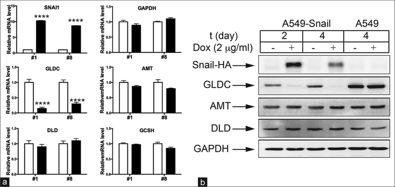
Figure 3:
Snail selectively suppresses the expression of glycine decarboxylase. (a) Two clones of A549‑Snail cells (clone #1 and #8) were grown in the absence (white bars) or presence (black bars) of 2 μg/ml Dox for 2 days. The levels of SNAI1, GAPDH, glycine decarboxylase, AMT, DLD, and GCSH mRNAs were measured using quantitative reverse transcription‑polymerase chain reaction. The result is expressed as fold change relative to the level of the uninduced control. (b) A549‑Snail or A549 parent cells were grown in the absence or presence of 2 μg/ml Dox for the indicated time. The levels of Snail‑HA, glycine decarboxylase, AMT, DLD, and GAPDH proteins were measured using Western blotting. ****P < 0.0001 (t‑test)

