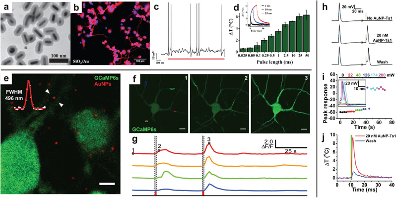
Figure 6.
Semiconductor nanoparticles can be excited by external light sources to stimulate growth and activity of neurons. a-d) Polymer-coated AuNRs excited by NIR resulted in neurite outgrowth. Current-clamp recording of a neuron showed action potentials fired in response to a single laser pulse, which is attributed to hyperthermal response of the AuNRs. Increased laser pulse produced an increased mean peak temperature change. Representative temperature profiles for various pulse lengths (inset). Reproduced with permission.[131] Copyright 2013, John Wiley & Sons. e-g) Fluorescent micrographs of Ca2+ concentration in neurons (green) and AuNPs (red). Representative images of varying Ca2+ concentration (green) in response to NIR excitation and corresponding spectral intensity response, illustrating the activation of neurons with NIR excitation at time points indicated by dashed lines. Reproduced with permission.[130] Copyright 2016, Nature Publishing Group. h-j) Functionalized AuNPs are robustly localized to neurons, and continue to stimulate neuronal activity after multiple washings. Increasing irradiance induces greater cell depolarization, triggering action potentials. The NIR-activated depolarization is attributed to the recorded, localized hyperthermia induced by excitation of the functionalized AuNPs. Reproduced with permission.[133] Copyright 2015, Cell Press.

