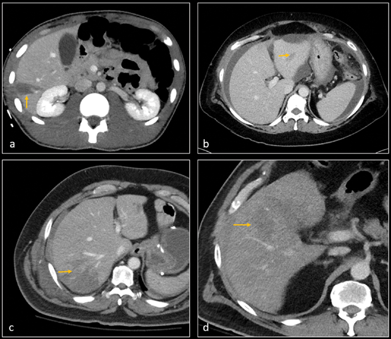
Fig. 1.

CT angiographies on four different patients show grade 2 liver lacerations (arrows) in segment 5 ( a ), segment 3 ( b ), segment 6 ( c ), and segment 4a ( d ). There was no evidence of active contrast extravasation and patients were hemodynamically stable. All patients remained stable and clinically improved with nonoperative management.
