
Fig. 3.
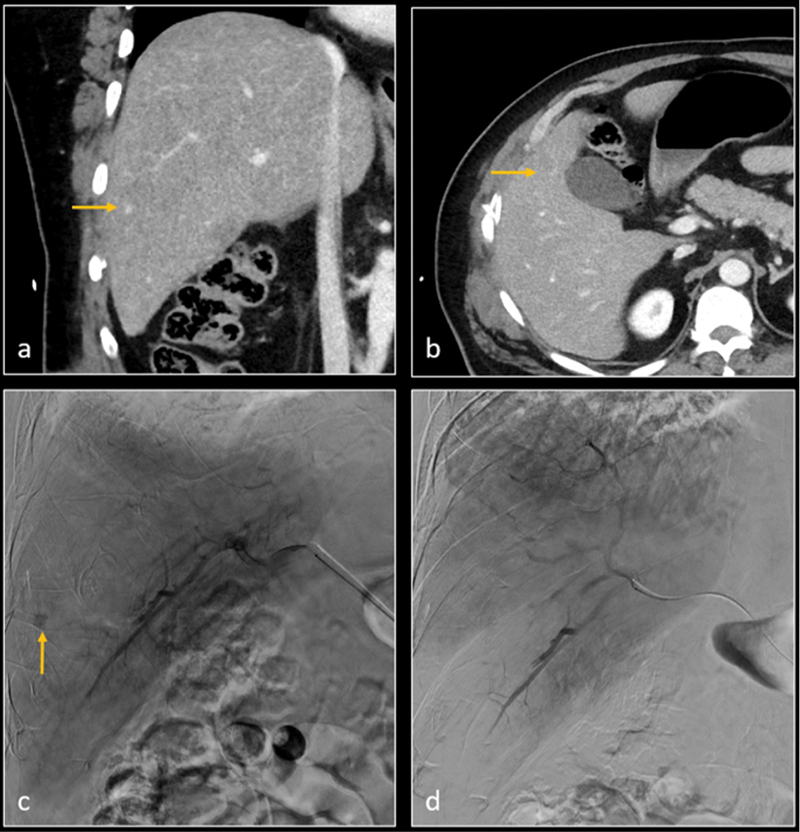
Coronal ( a ) and axial ( b ) CT images demonstrate hepatic segment 5 grade 2 laceration, and a small pseudoaneurysm (arrow). Digital subtraction angiography (DSA—image c ) confirms the pseudoaneurysm (arrow). Gelatin sponge embolization was performed. Subsequent DSA image ( d ) shows resolution of pseudoaneurysm.