
Fig. 6.
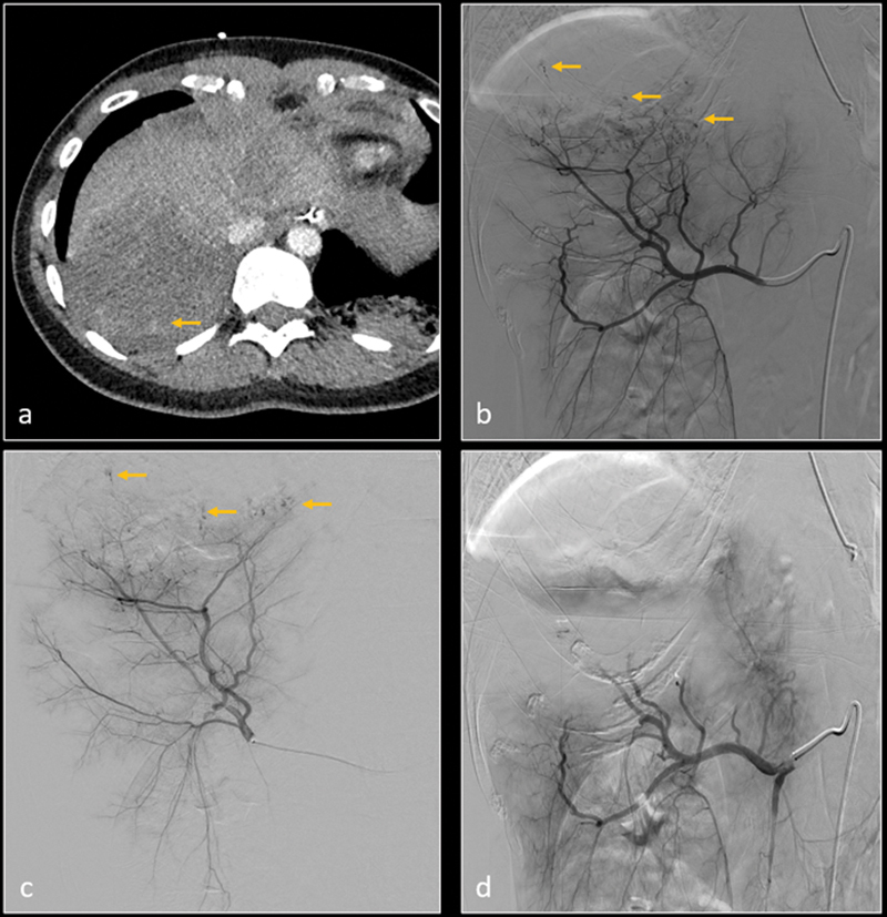
Axial CT image ( a ) shows grade 5 laceration in the right hepatic lobe with poor perfusion and small hemorrhagic foci (arrow). Subsequent digital subtraction angiography (DSA) of right hepatic artery ( b ) and posterior division of right hepatic artery ( c ) confirm multiple micro-hemorrhages (arrows). After gelatin sponge embolization, DSA of right hepatic artery ( d ) shows resolution of micro hemorrhages.