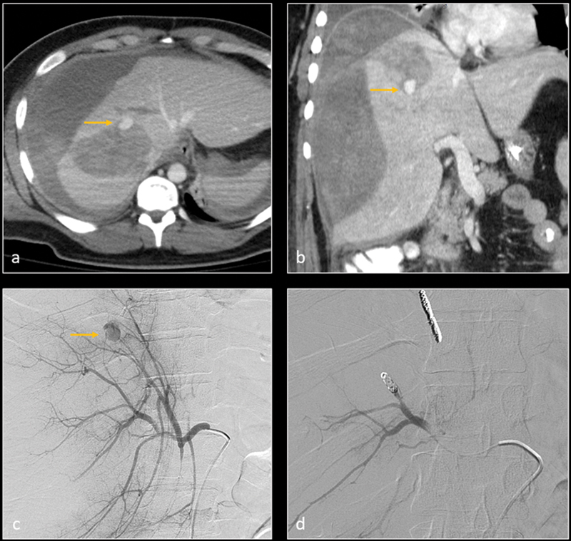
Fig. 7.

Axial ( a ) and coronal ( b ) CT images in arterial phase demonstrate grade 5 liver laceration with parenchymal hemorrhage, large subcapsular hematoma, and peritoneal hemorrhage. A small pseudoaneurysm vs. active extravasation is visualized within parenchymal hemorrhage (arrow). Digital subtraction angiography (DSA— c ) confirms the pseudoaneurysm (arrow). The feeding artery was embolized with metallic coils. Subsequent DSA image ( d ) shows resolution of the pseudoaneurysm.
