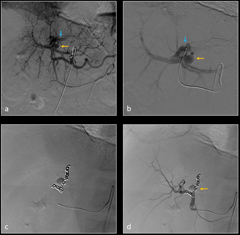
Fig. 8.

Patient was transferred from outside facility with no imaging studies available at the time of angiography. Digital subtraction angiography (DSA) of left hepatic artery ( a ) and more selective segment 2 hepatic artery ( b ) show a small left hepatic artery pseudoaneurysm (yellow arrow) and left hepatic artery–portal vein fistula (blue arrow). The feeding artery was selectively embolized using metallic coils ( c ). Subsequent DSA of left hepatic artery ( d ) demonstrates resolution of the pseudoaneurysm and arterioportal fistula.
