
Fig. 9.
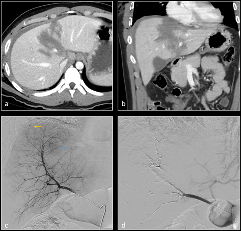
Axial ( a ) and coronal ( b ) CT images demonstrate grade 5 liver laceration. Selective digital subtraction angiography (DSA) of the right hepatic artery (RHA) anterior division demonstrates a small pseudoaneurysm (yellow arrow) and parenchymal blush (blue arrow). Selective embolization of the anterior division of RHA was performed with N-butyl cyanoacrylate (n-BCA) glue. Subsequent DSA image ( d ) shows resolution of the pseudoaneurysm and the suspected parenchymal hemorrhage.