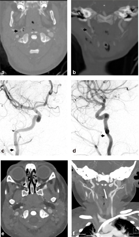
Fig. 1.

Biffl grade 1: A 22-month-old girl presented after multiple dog bites resulting in head, neck, and body injuries including skull and spine fractures. Axial ( a ) and coronal ( b ) computed tomography angiographic (CTA) images demonstrate small focal irregularity (arrows) of the distal right cervical internal carotid artery (ICA). Digital subtraction angiographic (DSA) images ( c —anteroposterior projection, d —lateral projection) demonstrate a Biffl grade 1 injury with focal narrowing (arrows) of the distal cervical right ICA near the skull base (arrows). Follow-up CTA ( e —axial, f —coronal images) 1 week later demonstrates resolution of the mild focal right ICA stenosis.
