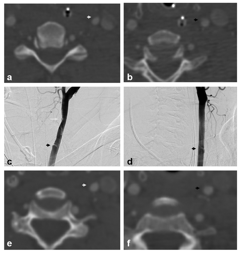
Fig. 2.

Biffl grade 2: A 55-year old presented after motor vehicle collision with cervical spine fracture and mild traumatic brain injury (TBI). Axial computed tomography angiographic (CTA) images demonstrate mild narrowing of the left common carotid artery (CCA) at C5 ( a , white arrow) and a dissection flap at C6/7 ( b , black arrow). Digital subtraction angiographic (DSA) images in oblique projections ( c, d ) demonstrate a Biffl grade 2 injury with focal narrowing of the left CCA at C5 (white arrow) and dissection flap at C6/7 (black arrows). Follow-up CTA axial images ( e, f ) 6 weeks later demonstrate improvement in vessel caliber at C5 (white arrow) and C6/7 (black arrow).
