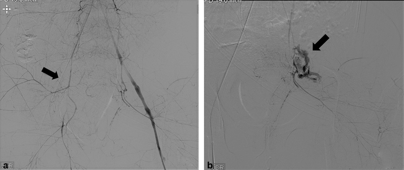
Fig. 1.

( a ) Digital subtraction angiography in a hypotensive patient with pelvic fracture shows that the right arterial access sheath is occlusive (arrow). Diffuse areas of spasm are seen without clear contrast areas of arterial injury. ( b ) Superselective catheterization of the left hypogastric artery demonstrates active extravasation near the origin of the inferior gluteal artery (arrow) that was later embolized with coils.
