
Fig. 18.
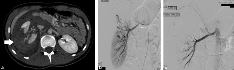
( a ) CT demonstrates a shattered right kidney with large perirenal hematoma (arrow). ( b ) Digital subtraction angiography (DSA) shows multiple areas of contrast extravasation in the mid and right upper pole. ( c ) After coil embolization of multiple branches, DSA shows preservation of vascular supply to the lower pole of the kidney.