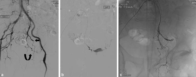
Fig. 19.

Pelvic fractures with hypotension. ( a ) Nonselective pelvic angiography shows transection of the left superior gluteal (arrow) and active extravasation from the inferior left gluteal artery (curve arrow). ( b ) Selective angiography redemonstrating active extravasation from the left inferior gluteal artery. ( c ) Angiography after coil embolization of both gluteal arteries demonstrates no further contrast extravasation.
