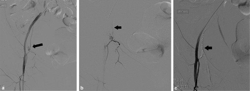
Fig. 4.

( a ) Digital subtraction angiography shows a pseudoaneurysm (PSA) near the origin of the inferior epigastric artery (arrow) in a patient with bleeding after an attempted femoral line. ( b ) Angiography shows coils placed distal to the PSA (arrow) to prevent retrograde filling through collaterals. ( c ) Postembolization angiography shows successful exclusion of the PSA using the sandwich technique and dense coil packing (arrow). As the lesion was close to the origin of the vessel, detachable coils were used.
