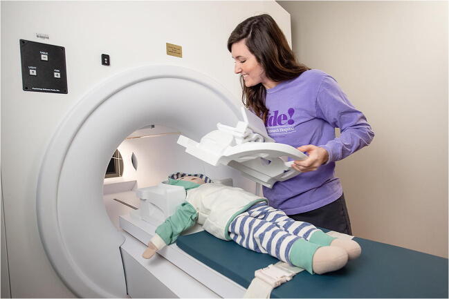
Fig. 4.
Mock MRI. Clinical image shows the mock MRI scanner, which has the same size bore as the majority of 1.5-T and 3-T scanners. A child life specialist demonstrates coil placement using a doll. After the process is completed, depending on preference, the child can choose to go through the mock experience and enter the scanner bore (image used with staff permission)

