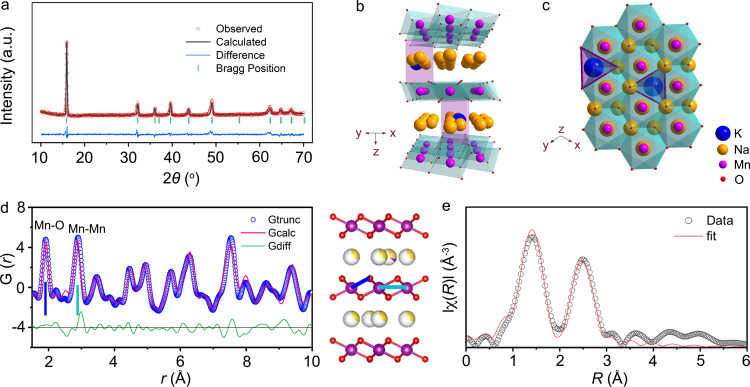
Fig. 1. Crystal structure of Na0.612K0.056MnO2.
a XRD pattern and Rietveld refinement. Typical layered structure of Na0.612K0.056MnO2 viewed along the y axis (b) and the z axis (c) with K+ located at the Nae sites. d PDF pattern and structure of Na0.612K0.056MnO2. The representative peaks correspond to the bond length of Mn–O (blue line) and Mn–Mn (pale blue line) as labeled. e Fitting of Mn K-edge FT-EXAFS spectra of Na0.612K0.056MnO2.

