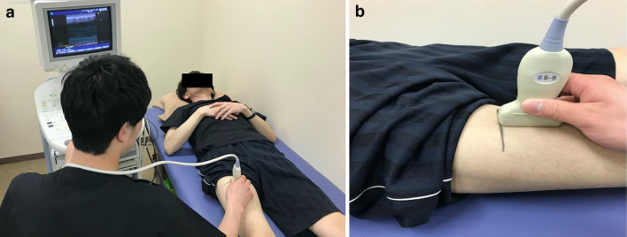
Fig. 1.
Ultrasonography set-up in the measurement of skeletal muscle thickness. a Participant is depicted in a supine position while the user holds the ultrasound probe against the anterior surface of the thigh. b The exact position of the probe is illustrated. Muscle thickness was determined from the resulting ultrasound images, and the values from two independent raters were used to decipher the intra- and inter-rater reliabilities

