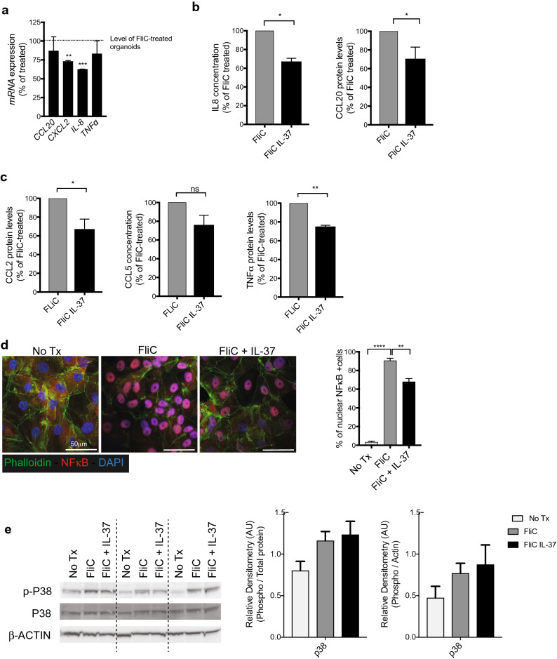
Figure 2.
IL-37 effect on human IEC FliC responses. (a) qPCR analysis of inflammatory genes after FliC and IL-37 exposure for 4 h expressed as percentage of change over FliC treated colonoids. (b) IL-8 and CCL20 protein levels (detected by ELISA) secreted basolaterally by human colonoids after 4 h of stimulation with FliC and IL-37. (c) CCL2, CCL5 and TNFa protein levels secreted basolaterally by colonoids after 4 h of stimulation with FliC and IL-37 (detected by Milliplex Luminex assay). (d) Immunostaining against NFkB (red), actin (Phalloidin—green) and DAPI (blue) of 2D monolayer after 30 min of stimulation with FliC with or without IL-37 (left). Counts of NFkB positive nuclei from immunostaining (right). (e) Western blot analysis of phospho and total p38, after 30 min of stimulation with FliC with or without IL-37 (left). Equal loading confirmed with β-Actin as well as total protein stain of membrane (see Fig. S4 online). Densities relative to total protein or β-Actin are shown (right). Mean and SEM are indicated from n = 4 donors (2 adult and 2 pediatric). All data shown are representative of at least 3 independent experiments. Statistical significance calculated using unpaired Student’s t-test *, P = 0.01 to 0.05; **, P = 0.001 to 0.01; ***, P = 0.0001 to 0.001; ****, P = 0.00001 to 0.0001; ns = not significant.

