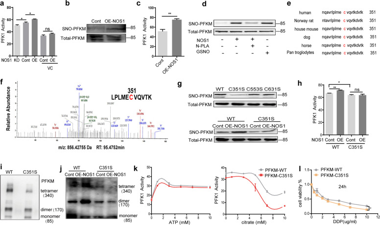
Fig. 3. NOS1 induced S-nitrosylation of PFKM at Cys351.
a The activities of PFK1 were measured in SKOV3-NOS1-KO, control, and SKOV3-OE-NOS1 cells vs. control and SKOV3-OE-NOS1 cells with ascorbic acid (VC) for 30 min (n = 3). b Western blot detected the content of SNO-PFKM protein in the control and OE-NOS1 tumor tissues which inoculated with SKOV3-OE-NOS1 and SKOV3-Cont cells. c The activities of PFK1 were measured in the SKOV3-control and SKOV3-OE-NOS1 tumor tissues (n = 3). d Biotin switch then purified all S-nitrosylated proteins in SKOV3 cells using streptavidin agarose resins. Western blot detected the content of S-nitrosation-modified PFKM (SNO-PFKM) protein in the SKOV3 cells, OE-NOS1 cells, the cells which were treated with GSNO (1 mM) for 30 min, and the OE-NOS1 cells treated with NOS1-specific inhibitor N-PLA (100 μM) for 48 h. e Amino acid sequence alignment of PFKM. The conserved cysteine residue (c, red) is S-nitrosated by NO. f Identification of the S-nitrosylation site on PFKM using mass spectrometry in SKOV3 cells. According to the data obtained by the mass spectrometer, the search database was searched by Thermo Proteome Discoverer 2.1. g Western blot detected the content of SNO-PFKM protein in SKOV3-PFKM-KO cells were transfected with Flag-PFKM WT or Flag-PFKM C351S, C553S, and C631S, respectively (upon). SKOV3-PFKM-KO cells and PFKM-KO+OENOS1 cells were transfected with Flag-PFKM-WT or Flag-PFKM-C351S, respectively. Western blotting detected the content of SNO-PFKM protein (down). h The enzymatic activity of PFK1 was detected in the SKOV3-PFKM-KO cells and PFKM-KO+OENOS1 cells which were respectively reconstituted with Flag-PFKM-WT or Flag-PFKM-C351S. i Western blot detected the contents of PFKM tetramer, dimer, and monomer in the SKOV3-PFKM-KO cells which were respectively reconstituted with Flag-PFKM-WT or Flag-PFKM-C351S. j Western blot detected the changes in PFKM tetramer, dimer, and monomer in the SKOV3-PFKM-KO cells and PFKM-KO+OE-NOS1 cells which were respectively reconstituted with Flag-PFKM-WT or Flag-PFKM-C351S. k Different concentrations of ATP (0–10 mM, left) or citrate (0–10 mM, right) were added to observe the activity of PFK1 from the total protein in SKOV3-PFKM-WT or SKOV3-PFKM-C351S cells (n = 3). l Cell viability% detected by CCK8 assay. Different concentrations of DDP were applied for 24 h in the SKOV3-PFKM-WT and SKOV3-PFKM-C351S cells, respectively (*P < 0.05 by one-way ANOVA).

