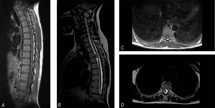
Fig. 1. Simple dorsal MRI.
Right: A sagittal section shows an extra-axial mass at T3-T4, isointense T1 (a) and hypointense T2 signals (b), and displacement of the spinal cord posteriorly. Left: Axial section showing a T1 hypointense left ventrolateral mass (c) and a T2 hyperintense mass (d) displacing the spinal cord to the right and posteriorly.

