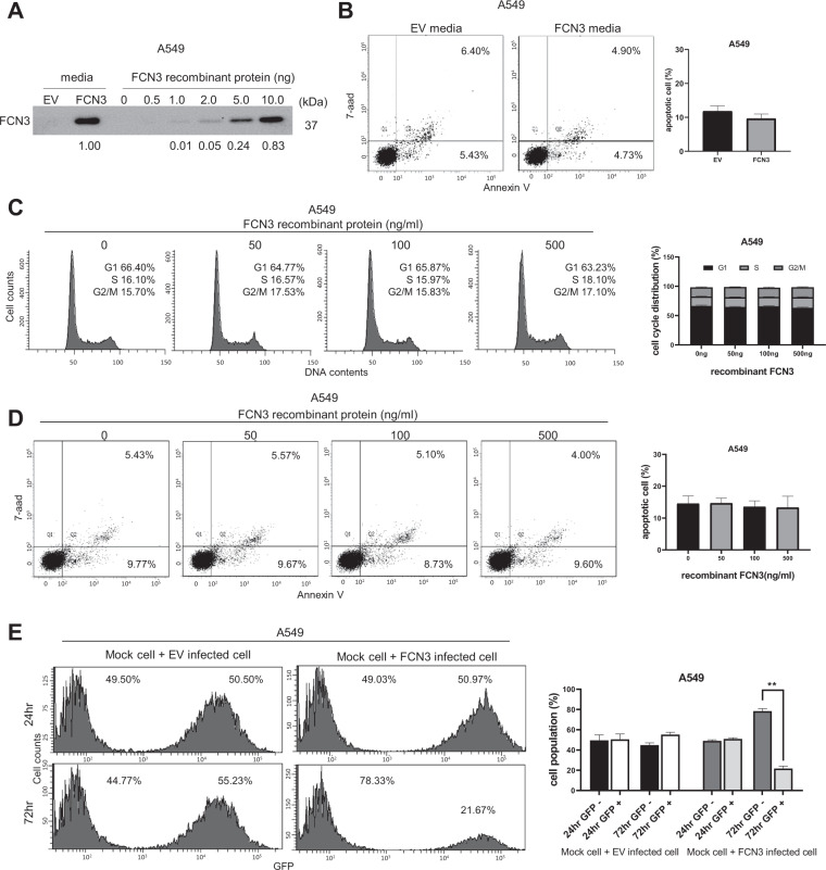
Fig. 4. Tumor suppressor activity of FCN3 involves an intracellular mechanism.
A Immunoblot showing the secreted FCN3 in cultured media of FCN3-virus-transduced A549 cells but not in culture media of control virus (EV)-transduced cells. Indicated amounts of recombinant FCN3 protein are also shown. B Culture media from control virus- or FCN3-virus-transduced cells were applied to A549 cells, and apoptosis was analyzed by flow cytometry after 96 h. Data summarized in the graph are mean ± SEM of three independent experiments. Note no significant difference was seen. C Cell cycle analyses of A549 cells by flow cytometry 72 h after application of indicated doses recombinant FCN3. No alteration in cell cycle progression was observed. Data are mean ± SEM of three independent experiments. D Apoptosis was examined 96 h after application of indicated doses recombinant FCN3. No alteration in proportions of apoptotic cells was noticed. Data are mean ± SEM of three independent experiments. E A549 cells with or without viral transduction were mixed and co-cultured for 24 h and 72 h. Proportions of GFP-negative and -positive cell populations were analyzed by flow cytometry. After 72 h, GFP-positive proportion was decreased only in the case of FCN3-virus-transduced cells. Data are mean ± SEM of three independent experiments, and (**) represents P-value of <0.01 from t test.

