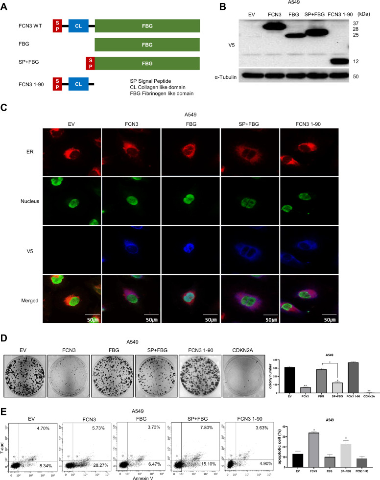
Fig. 6. The FBG domain localized to ER is necessary and sufficient for tumor suppressor activity of FCN3.
A Schematic illustration of various FCN3 derivatives: wild type FCN3, FBG domain, SP (signal peptide) + FBG domain, and FCN3 1-90. B Immunoblots showing ectopic expression of various FCN3 derivatives in A549 cells. Antibody against V5 epitope was used. ɑ–Tubulin was used as the loading control. C Expression and subcellular localization of FCN3 derivatives in A549 cells. Nuclear GFP expression indicates the virus-transduced cell. V5 staining shows localization of various FCN3 derivatives. Note no V5 staining in control virus (EV)-transduced cells and nuclear localization of FBG. D Colony formation assay after transducing with control virus or viruses expressing the indicated FCN3 derivatives in A549 cells. Note SP + FBG has growth inhibition effect. Numbers of colonies were counted and presented in bar graphs. Data are mean ± SEM of three independent experiments. E Apoptosis of A549 cells was evaluated with flow cytometry after transducing with control virus or viruses expressing the indicated FCN3 derivatives. (*) and (**) represent P-values of <0.05 and <0.01 from t tests, respectively.

