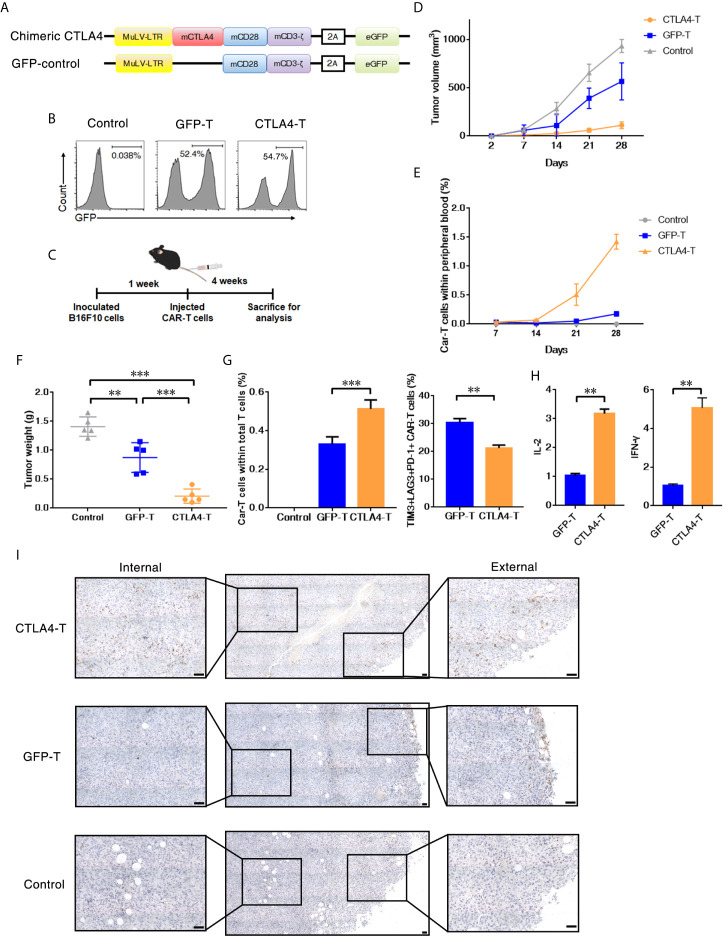
Figure 3.
T cells expressing the CTLA4-CD28-CD3z chimera had effective tumor infiltration. (A) Murine chimeric CTLA4 molecules contained the extracellular and transmembrane domains of mouse CTLA4, the cytoplasmic region of mouse CD28, and the intracellular domains of mouse CD3z. T cells expressing GFP were constructed as the control group. (B) Representative flow cytometric analysis of murine chimeric CTLA4 or GFP expression in mouse T cells. (C) Experimental scheme for evaluating murine CTLA4-CAR T cells efficacy, 2 × 10⁵ of B16F10 cells were subcutaneously transplanted, and mice were intravenously administered T cells transduced with either chimeric CTLA4 or GFP or PBS (Control), five mice/group. (D) The tumor volumes in the mice were measured and calculated every 7 days. (E) The percentages of CAR T cells in peripheral blood of the mice were measured and calculated every 7 days. (F) The B16F10 tumor weight was weighed after 35 days, mean ± SD, one-way ANOVA. (G) The percentages of CAR T cells in total infiltrated T cells within the tumor tissues, and the percentages of TIM3+LAG+PD-1+ CAR T cells, mean ± SD, one-way ANOVA. (H) qRT-PCR analysis of the mRNA expression of the indicated genes. The results were normalized to glyceraldehyde 3-phosphate dehydrogenase (GAPDH) mRNA levels and are presented as the mean ± SEM (n = 3), unpaired two-tailed t-test. (I) Immunohistochemical staining identified the infiltrated CAR T cells in resected tumors, GFP+ cells were stained. Significance values: **P < 0.01; ***P < 0.001.

