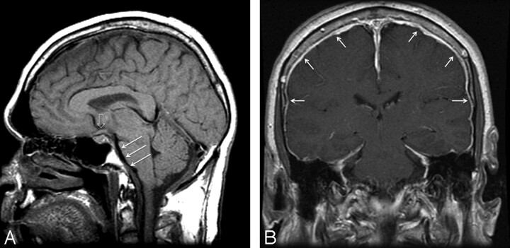
Fig 1.
Classic findings of SIH on MR imaging of the brain with “brain sag” demonstrated on midline sagittal T1 image (A), including descent of the cerebellar tonsils below the foramen magnum, flattening of the ventral pons (white arrows), and inferior displacement of the optic chiasm (open arrow). Postgadolinium coronal T1 image (B) demonstrates diffuse dural enhancement (white arrows.)

