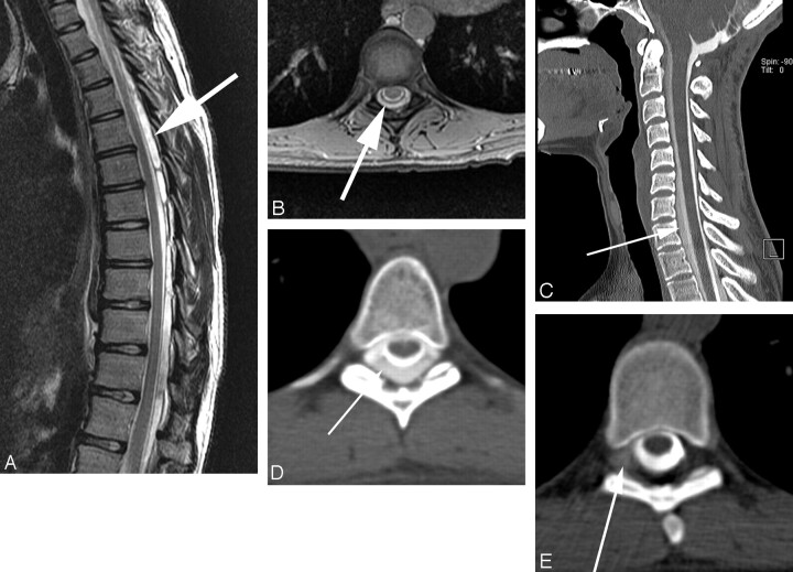
Fig 2.
Fast CSF leak in a patient with extra-arachnoid fluid on spinal MR imaging, localized on dynamic CTM. Large extra-arachnoid fluid collection is seen on sagittal T2 images of the thoracic spine, predominantly posterior to the thecal sac (A), which involved the entire cervical and thoracic spine. Axial gradient images through the spine confirm extra-arachnoid fluid (B). Conventional CTM shows a collection of contrast outside of the thecal sac (C, D), which extends from C3 to L4 and the leak cannot be localized. Dynamic CTM shows extraarachnoid extravasation of contrast on the right at T6-T7 (E) and also at right T7-T8 (not shown), consistent with 2 sites of CSF leak.

