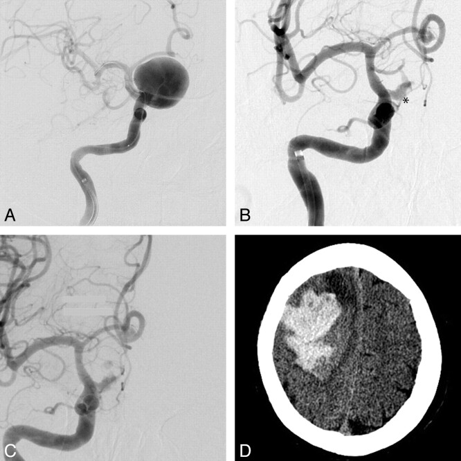
Fig 3.
A, Initial right ICA anteroposterior (AP) angiogram with a giant supraclinoid ICA aneurysm before PED deployment (the stent is collapsed within the microcatheter). Right ICA AP angiogram 3 months later, with persistent early aneurysm filling (asterisk) pre- (B) and postdeployment (C) of 2 more PEDs. The aneurysm sac showed significant interval enlargement and development of a daughter bleb (images not shown), so a new PED was placed to increase the flow effect. D, NCCT 1 day later shows a large acute right lobar hematoma with significant mass effect. The patient survived with a persistent left attenuated hemiparesis.

