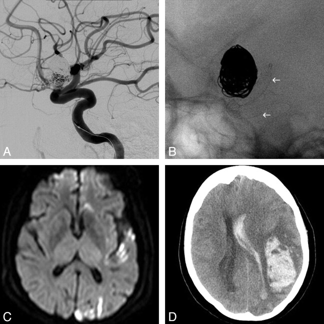
Fig 4.
A, Left ICA lateral angiogram showing the previously coiled supraclinoid aneurysm with a significant residual filling. B, Single-shot lateral image with the PED fully deployed covering the neck of the aneurysm (arrows). C, DWI 48 hours later shows multiple left cortical ischemic lesions. D, NCCT 6 days later shows a large left frontal lobar hematoma, emptying into the ventricle, with marked midline shift and mass effect. The patient died 24 hours late.

