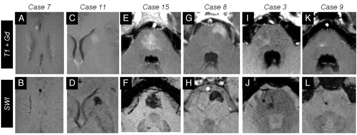
Fig 2.
Six representative cases of BCTs on gadolinium-enhanced (upper row) and SWI images (lower row). A–D, Cases 7 and 11 show supratentorial BCTs. Notice the signal intensity drop on SWI in case 7 (B) and the BCT in the head of caudate in case 11 (C). Four cases of infratentorial BCTs are presented. E–H, Cases 15 and 8 present rather large BCTs: a central pontine BCT with irregular brushlike borders (E and F) and a superficial, left ventrolateral pontine BCT in case 8 (G and H). I–L, Cases 3 and 9 are smaller BCTs of the pons. Note that SWI more clearly depicts the prominent vessel pointing to the BCT.

