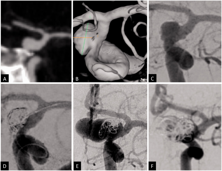
Figure 2.
Coronal CT angiogram (a), 3D volume-rendered angiogram reconstruction (b) and 2D anteroposterior angiogram (c) showing a ruptured 8 × 5 mm posterior communicating artery aneurysm with 5 mm neck. The aneurysm was treated with balloon assisted coiling. A small remnant was left at the inferior neck due to difficulty of recannulating this pocket (d). The plan was for elective flow diversion after the patient’s acute hospitalization phase. While recovering in the rehabilitation center, the patient developed transient right face and arm weakness 1 month after her initial hemorrhage. CT angiogram revealed major recurrence of the coiled aneurysm, measuring approximately 17 × 9 mm with multiple lobulations that was confirmed by angiography (e). The patient was retreated with coils and flow diversion across the aneurysm (d).

