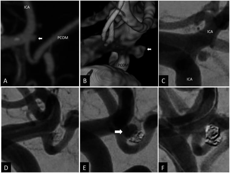
Figure 5.
Sagittal CT angiogram (a), 3D volume-rendered angiogram reconstruction (b) and 2D oblique angiogram (c) showing a ruptured 2 × 2 mm left posterior communicating artery aneurysm (white arrow in (a) and (b)). The aneurysm was coiled using balloon assisted coiling with satisfactory occlusion (d). Routine cerebral angiogram done at 20 weeks showed recurrent 4 × 3 mm posterior communicating artery aneurysm (white arrow) with a 1.2 mm neck (e) that was retreated with balloon assisted coiling (f).

