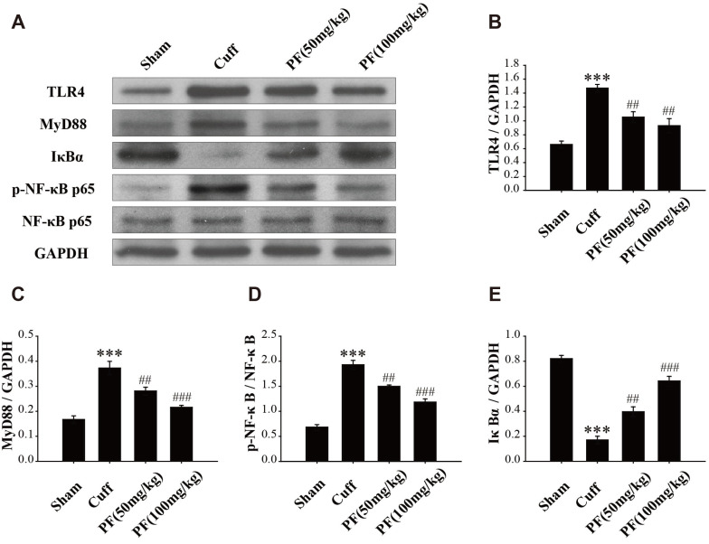
Fig. 6. PF inhibits the increase in expression of TLR4/NF-κB pathway induced by Cuff.
The expression of TLR4, MyD88, IκBα, p-NF-κBp65, and NF-κBp65 were determined by western blot analysis in the hippocampus (A). Statistical analysis of relative levels of TLR4 (B), MyD88 (C), p-NF-κBp65 (D), and IκBα (E). Quantitative results are expressed as mean ± standard deviation. n = 3. PF, paeoniflorin. ***p < 0.001 vs. Sham group; ##p < 0.01, ###p < 0.001 vs. Cuff group.

