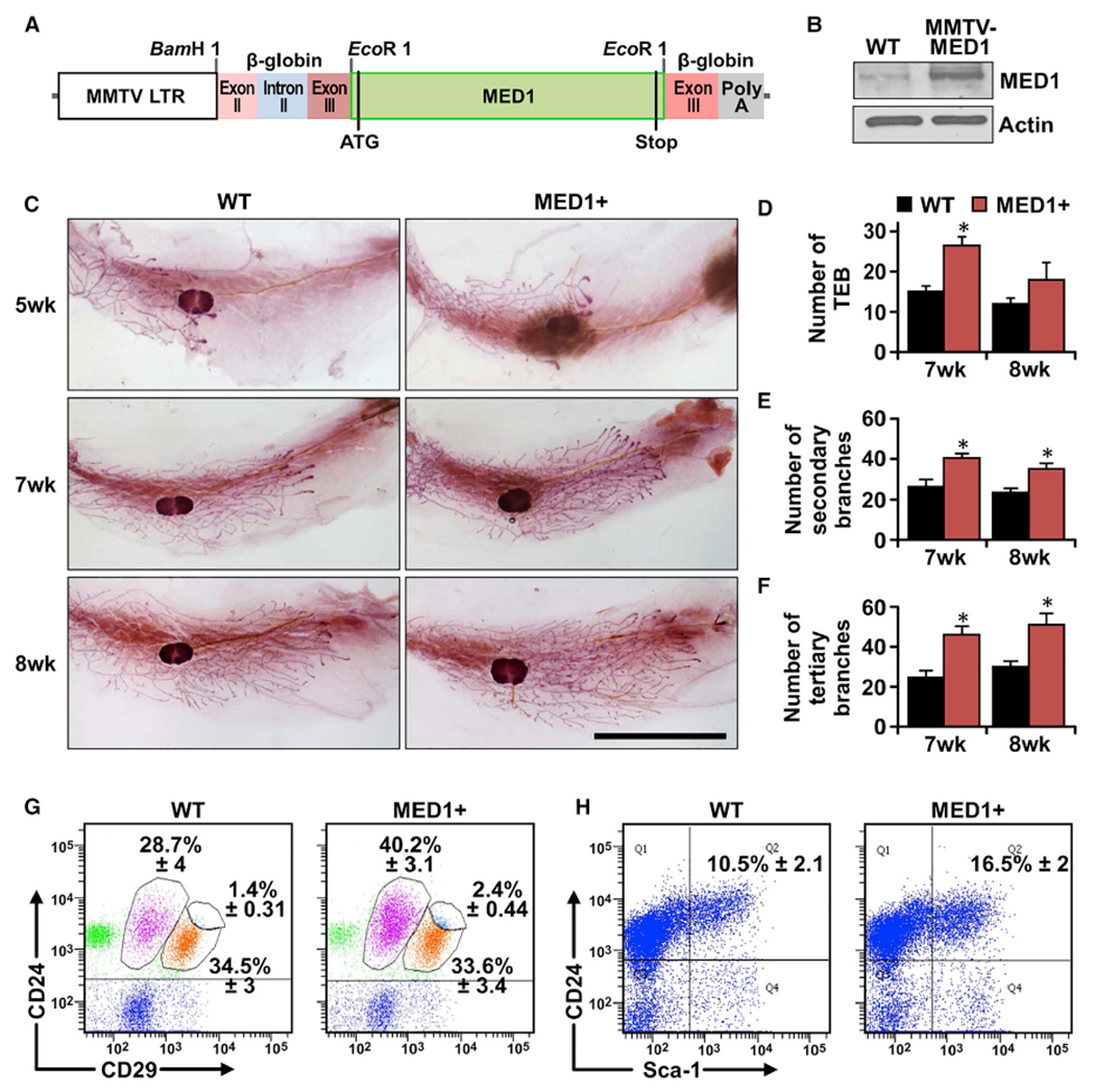
Figure 1. Generation and characterization of mammary-gland-specific MED1-overexpression mice.

(A) Schematic representation of MED1-overexpression transgenic construct (MMTV-MED1).
(B) MED1 expression in mammary epithelial cells of MMTV-MED1 transgenic mouse was assessed by immunoblotting.
(C) Whole-mount staining of pubertal mammary gland at age of 5, 7, and 8 weeks. Scale bar: 1 cm.
(D–F) Analyses of the number of terminal end buds (TEBs) (D), secondary branches (E), and tertiary branches (F) of mice in (C).
(G) Mammary cells from 7-week-old wild-type (WT) and MMTV-MED1 mice were analyzed by flow cytometry using antibodies against cell surface markers Lin (CD31, CD45, and Ter119), CD24, and CD29.
(H) Flow cytometry analyses of Lin−CD24+Sca1+ cells in mammary glands of 7-week-old WT and MMTV-MED1 mice. The values are obtained from three independent experiments and shown as mean ± SD. *p < 0.05.
