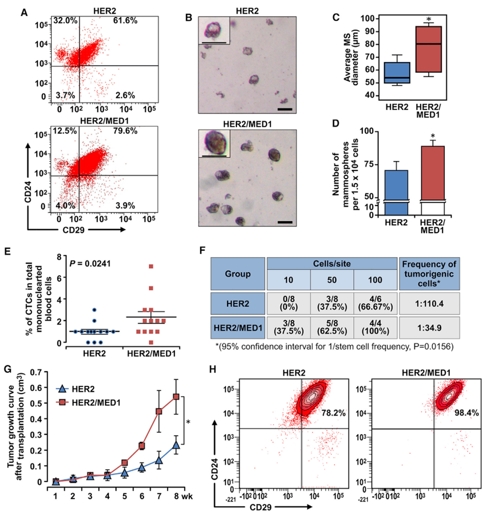
Figure 4. MED1 overexpression promotes MMTV-HER2 CSC formation.

(A) FACS analyses of MMTV-HER2 and MMTV-HER2/MMTV-MED1 CSCs using antibodies against cell surface markers Lin, CD24, and CD29.
(B) Mammosphere assays using FACS-sorted tumor cells in (A). Scale bar: 100 μm.
(C and D) Average diameters (C) and numbers (D) of mammospheres formed in (B).
(E) Statistics of flow cytometry analysis of CD45−CK18+EpCAMhi circulating tumor cells (CTCs) in mononuclear blood cells from MMTV-HER2 and MMTV-HER2/MMTV-MED1 tumor-bearing mice (n = 13).
(F) Limiting dilution analyses of tumor-initiating cells in MMTV-HER2 and MMTV-HER2/MMTV-MED1 bulk tumors.
(G) Growth curves of orthotopic MMTV-HER2 and MMTV-HER2/MMTV-MED1 tumor xenografts (n = 6).
(H) FACS analyses of the grafted tumors using cell surface markers Lin (CD31, CD45, and Ter119), CD24, and CD29. The values are obtained from three independent experiments and shown as mean ± SD. *p < 0.05.
