Figure 2.
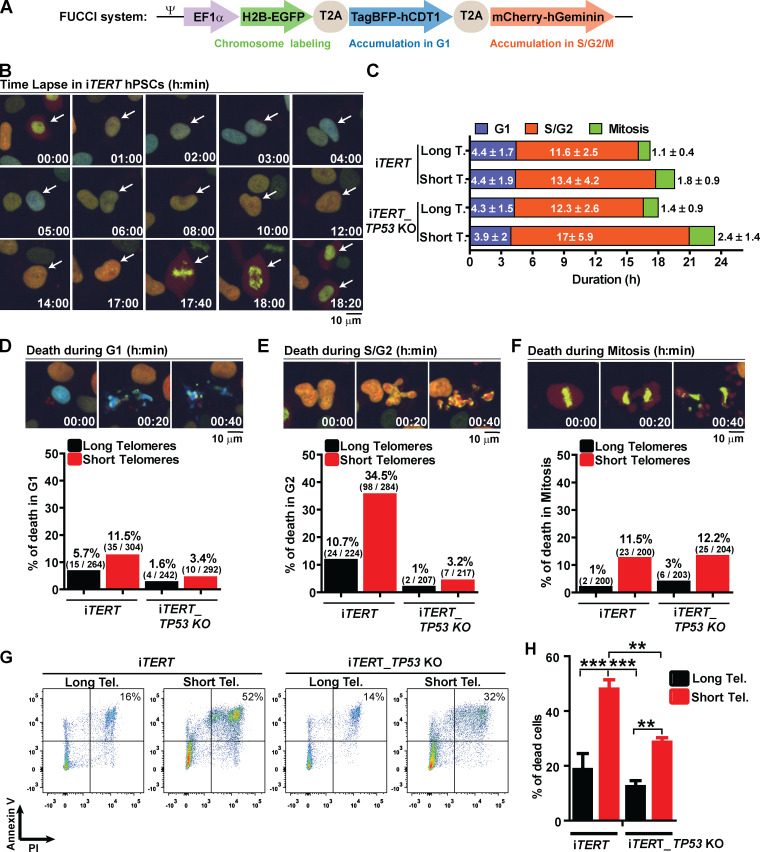
Telomere attrition prolongs S/G2 and mitosis duration in hPSCs, leading to p53-mediated cell death. (A) FUCCI system: Cells express EGFP-H2B (green fluorescence to track mitotic chromosomes), TagBFP (blue fluorescence in G1), and mCherry (red fluorescence in S/G2/M). (B) Representative time-lapse image of FUCCI live imaging. (C) Average duration (h) ± SEM of 100 events analyzed per phase (G1, S/G2, or M) per cell. (D–F) Illustrative images and percentage cell death during G1, S/G2, or mitosis, respectively. At least 200 events were analyzed per cell cycle phase per cell and scored as live (able to transit to the next cell cycle phase) or dead (died within that specific cell cycle phase). (G and H) Cell death analysis by Annexin V and PI staining. Average ± SD of three independent experiments is shown in H. Tel., telomere. Statistical analysis: one-way ANOVA followed by Bonferroni’s test. **, P < 0.01; ***, P < 0.001. PDLs for cells with short telomeres: 159 (iTERT hPSCs); 209 (iTERT_TP53 KO hPSCs).

