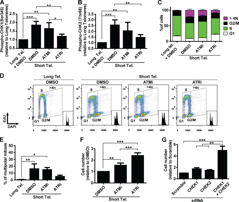
Figure 4.
A determinant role for ATR after telomere (Tel.) attrition in hPSCs. (A and B) Analysis of phosphoCHK1 (Ser345) and phosphoCHK2 (Thr68), respectively, by flow cytometry in iTERT hPSCs with short telomeres in the presence of ATMi (10 µm) or ATRi (1 µm). Average ± SD of five independent experiments. (C and D) Cell cycle distribution in iTERT hPSCs with short telomeres treated with ATMi or ATRi. Average ± SD of three independent experiments are shown in C and representative images in D. (E) Multipolar mitosis analysis of iTERT hPSCs with short telomeres in the presence of ATMi or ATRi by immunofluorescence. (F) Cell proliferation analysis in iTERT hPSCs with short telomeres in the presence of ATMi or ATRi. (G) Cell proliferation analysis in iTERT hPSCs with short telomeres 7 d after transfection with the indicated siRNAs. In E–G, average ± SD of three independent experiments. Statistical analysis: one-way ANOVA followed by Bonferroni’s test. *, P < 0.05; **, P < 0.01; ***, P < 0.001. PDLs for iTERT hPSCs with short telomeres: 149–159.

