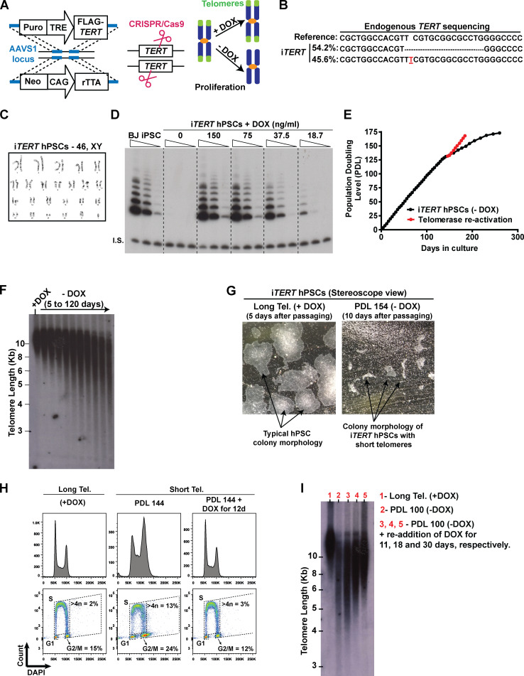
Figure S1.
Establishing a novel model to study telomere (Tel.) dysfunction in hPSCs. (A) Engineering iTERT hPSCs. First, TRE-Tight–regulated FLAG-TERT cDNA and CAG-driven rTTA sequences were inserted into the AAVS1 safe harbor locus of BJ-iPSCs using zinc fingers. After clonal selection, the endogenous TERT gene was knocked out using CRISPR/Cas9. Telomerase activity in the resulting cell (iTERT hPSC) is regulated by DOX. (B) NGS confirming the deletion of the TERT endogenous locus. (C) Karyotype analysis of iTERT hPSCs. (D) TRAP analysis of iTERT hPSCs treated with different concentrations of DOX. Unedited BJ-iPSCs were used as positive control. I.S., internal standard. (E) PDLs of iTERT hPSCs grown in the absence of DOX (black curve) or after telomerase reactivation (red curve). (F) Telomere length analysis by TRF in iTERT hPSCs kept with or without DOX for the indicated time. (G) Representative images of iTERT hPSCs colonies grown in the presence of DOX (long telomeres) or at PDL 154 after DOX withdrawal (short telomeres). (H) Readdition of DOX for 12 d fully rescued the cell cycle profile of iTERT hPSCs as detected by EdU and DAPI staining (flow cytometry). (I) Telomere length analysis by TRF in iTERT hPSCs grown continuously with DOX (long telomeres, lane 1), with short telomeres (PDL 100, lane 2), and after readdition of DOX for 11, 18, or 30 d (lanes 3, 4, and 5, respectively) to the culture media of the cells with short telomeres.

