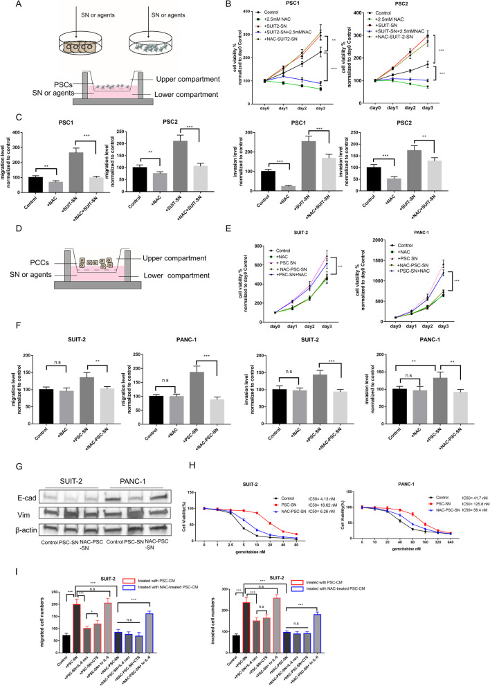
Fig. 3.
NAC attenuates cancer-stroma interaction in pancreatic cancer. a The schema for indirect co-culture experiments to evaluate the effects of NAC between PCCs and PSCs. (top) The supernatant (SN) was obtained as described above and then added as CM to the following experiments as indicated. (bottom) Effects of NAC in PSCs which induced by PCC-SN in the co-culture system. PCC-SN or agents were added to the plate, and PSCs were seeded in upper Chambers (8 μm). The number of migrated or invaded (c) PSCs were evaluated after incubation for 24 h or 48 h. b Cell viability assays of PSCs co-cultured with various agents. PSCs were not treated (Control) or added with 2.5 mM NAC, or SUIT2-SN, or SN from NAC-treated SUIT-2(NAC-SUIT2-SN), or dual treated with SUIT2-SN and NAC. c Migration and invasion assays of PSCs were performed as described in A (bottom) for 24 h or 48 h, respectively. Migrated or invaded cell numbers were normalized by the total cell number of each cell. For details, also see supplementary figure S4A. d The schema for effects of NAC in PCCs which induced by PSC-SN in the co-culture system. PSC-SN or agents were added to the plate, and PCCs were seeded in upper Chambers (8 μm). The number of migrated or invaded (F, I) PCCs were evaluated after incubation for 24 h or 48 h. e Cell viability assays of PCCs co-cultured with various agents. PCCs were not treated (Control) or added with 5 mM NAC, or PSC-SN, or SN from NAC-treated PSC (NAC-PSC-SN), or dual treated with PSC-SN and NAC. f Migration and invasion assays of PCCs were performed as described in D for 24 h or 48 h, respectively. Migrated or invaded cell numbers were normalized by the total cell number of each cell. For details also see supplementary figure S5. g Expression of E-cadherin and Vimentin in PCCS treated with PSC-SN or NAC-PSC-SN by western blotting. h Cell viability assays of PCCs at different concentrations of Gemcitabine after 72 h treatment. PCC cells were pre-processed with PSC-SN or NAC-PSC-SN for 48 h. Cell viability in all groups was normalized to their viability treated with 0 nM Gemcitabine. i Migration and invasion assays of SUIT-2 cells were performed at 24 h or 48 h after treatment with indicated agents. Migrated and invaded cells were counted. Agents were as followed: SN from PSC (PSC-SN), 5 μg/ml IL-6 neutralizing antibody (IL6 neu), 20 μM Cryptotanshinone (CTS), 20 ng/ml human recombination IL-6 (hr IL-6), SN from NAC-treated PSC (NAC-PSC-SN). Also, see supplementary figure S5C for details and figure S5D for assays of PANC-1 cells. *P < 0.05; **P < 0.01; ***P < 0.001; n.s, no significance

