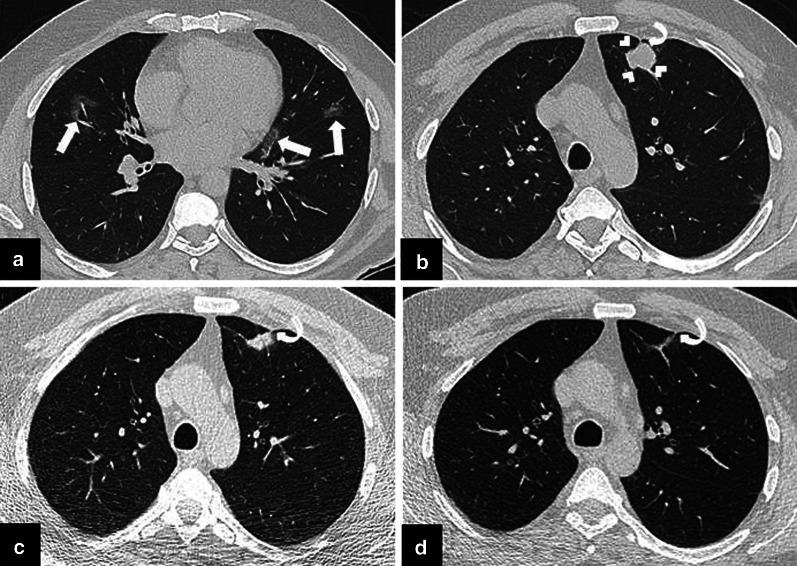
Fig. 1.
a, b The initial chest CT scan obtained following PCR test positivity for COVID-19 infection, revealed a few patchy areas of ground glass opacity (GGO) in both lungs (arrows) compatible with COVID-19 pneumonia. An irregularly shaped solid nodule 2 cm in diameter in left upper lobe of the lung was also noted (arrowheads). Percutaneous transthoracic core needle biopsy was scheduled due to suspicion of primary lung cancer. c CT scan obtained prior to biopsy procedure demonstrated significant size reduction of the nodule. Therefore, biopsy was not performed. d Follow-up CT scan obtained 3 months later demonstrated complete resolution of the nodule. A pleural tag which became more apparent following resolution of the nodule (curved arrows, b–d) raised the suspicion of COVID-19 triggered focal organizing pneumonia

