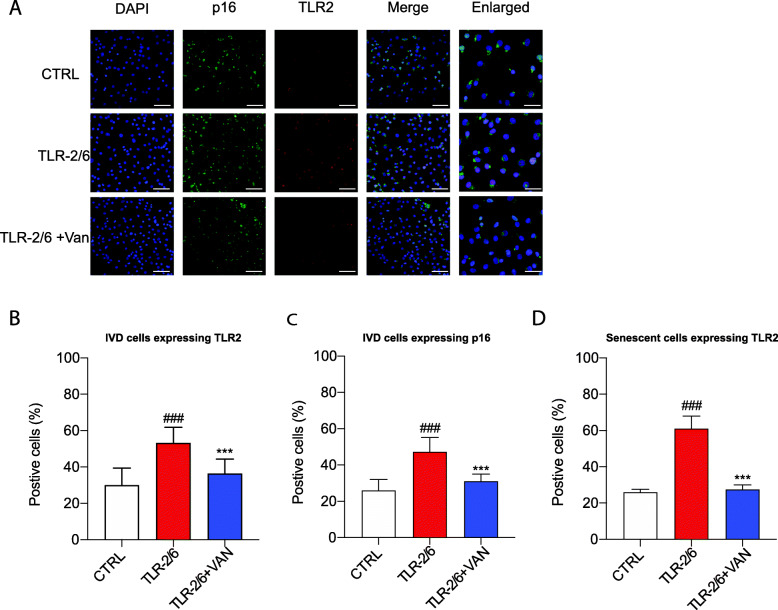
Fig. 5.
o-Vanillin reduced the number of cells co-expressing TLR-2 and p16ink4a in cells exposed to TLR-2/6 agonist. Disc cells from degenerate IVDs were induced with TLR-2/6 agonist for 48 h with (TLR-2/6 + VAN) or without (TLR-2/6) o-vanillin treatment for 6 h or no induction with TLR-2/6 agonist (CTRL). a Photomicrographs of IVD cells stained for DAPI (blue) and either p16ink4a (green), TLR-2 (red), or the merge (p16ink4a and TLR-2) as revealed by Immunocytochemistry. DAPI, p16, TLR2, and merge images scale bars = 25 μm. Enlarged images scale bar: 10 μm. b–d Quantification of the percentage of IVD cells that stained positive for b TLR-2, c p16ink4a, or d co-localized cell for TLR-2 and p16ink4a. Percentage of positive cells in e were the average for n = 5. #p < 0.05, ##p < 0.01, and ###p < 0.001 indicate significant difference between the TLR-2/6 agonist treated to the non-induced control and *p < 0.05, **p < 0.01, and ***p < 0.001 indicate significant difference between the TLR-2/6 agonist with o-vanillin treated to the TLR-2/6 agonist treated. b–d Mean ± SD, statistical analysis was done using paired t-test

