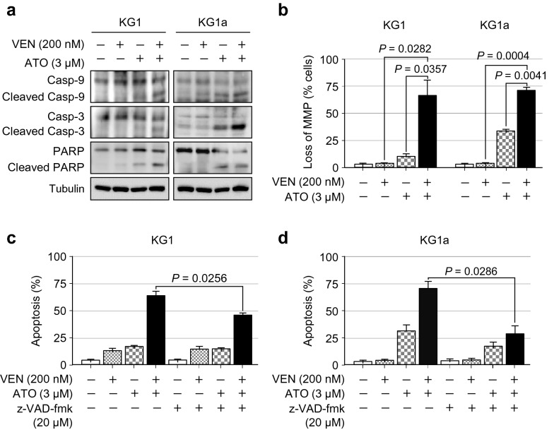
Fig. 4.
The combination of venetoclax and ATO promotes caspase-dependent apoptosis in KG1 and KG1a cells. a, Representative western blot analysis of the indicated proteins in KG1 and KG1a cells treated with venetoclax (200 nM), ATO (3 µM), or both in combination for 48 h. Similar results were obtained from three independent experiments. b, Summary data of MMP disruption, as assessed by flow cytometry, using the DePsipher Kit (Trevigen) in KG1 and KG1a cells treated with venetoclax (200 nM), ATO (3 µM), or both in combination for 48 h. Values were obtained from three independent experiments, and horizontal bars indicate mean ± s.d. *P values versus control treatment by two-tailed Mann–Whitney U test. n.s., not significant. c, d, Summary data of the percentage of apoptotic cells, as assessed by Annexin V/PI staining and flow cytometric analysis after treatment with or without the pan-caspase inhibitor z-VAD-fmk (20 µM) for 2 h prior to the addition of venetoclax (200 nM), ATO (3 µM), or both in combination for 48 h in KG1 (c) and KG1a (d) cells. Values were obtained from three independent experiments, and horizontal bars indicate mean ± s.d. *P values versus control treatment with or without z-VAD-fmk by two-tailed Mann–Whitney U test. n.s., not significant

