
Figure 4.
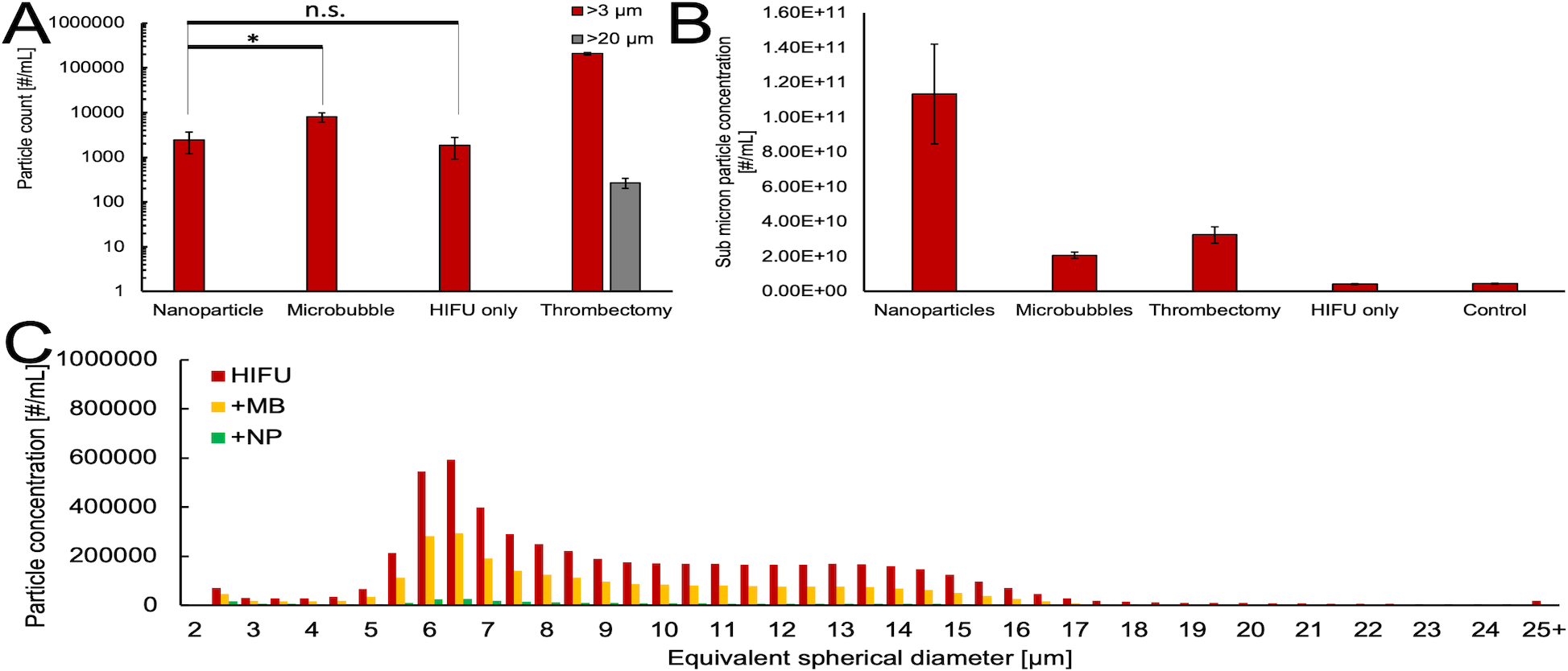
A) Counts for >3 and >20 μm equivalent spherical diameter particles from FLOWCAM imaging for each treatment. *Indicates significant difference at α=0.05, one-sided; n.s. denotes non-significance. B) Submicron (<1 μm) particle counts as estimated from Nanoparticle Tracking Analysis, with background subtracted. The error bars correspond to one standard deviation. C) FLOWCAM particle histograms for various treatments applied to the supernatant of a thrombus after mechanical thrombectomy condition was applied. The histograms have a 0.5 μm bin size and are an amalgamation of four independently treated samples.